Anatomy Diagram Rib Area : Human Rib Cage Anatomy Stock Illustration 65588847 Pixta. In this image, you will find thoracic vertebrum, costochondral joint, costal cartilage, costal margin, costal arch, thoracic vertebrum, xiphoid process, xiphisternal joint, body, manubrial sternal joint, manubrium, the sternal notch in it. The primary responsibilities of the ribcage involve protecting the thoracic visceral organs, enclosing the thoracic visceral organs, and is included. The head only articulates with the body of the t1 vertebra and therefore only one articulatory surface is present. Anatomy of the rib cage diagram. Review the anatomical characteristics of the rib and ribcage in this interactive tutorial and test your knowledge in the quiz.
A typical human rib cage consists of 24 ribs in 12 pairs the sternum and xiphoid process the costal cartilages and the 12 thoracic vertebrae. They make up the lateral part of our body, its anterior and posterior wall and they entirely build the lateral parts of the chest wall. Anatomy of the rib cage diagram. In comparison, the first two ribs are shorter and more curved. Our latest youtube film is ready to run.
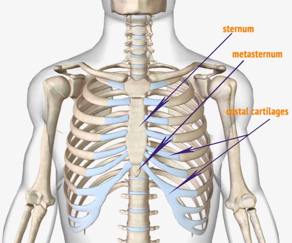
Https Encrypted Tbn0 Gstatic Com Images Q Tbn And9gcsbcwlitzqg 4gi75sywgmufbxcsx5tgikkiaqgzv Eslsk5gyh Usqp Cau from The major muscles of the abdomen include the rectus. The heads of ribs 1, 10, 11, and 12 have a single facet for articulation with the bodies of the thoracic vertebrae. Several muscles that move the arms, head, and neck have their origins on the sternum. Anatomy diagram rib area : Learn about lung function, problems, location in the body, and more. These muscles help the body bend at the waist. The anatomy of the human ribs is made up of 24 ribs which are parted in 12 pairs (each on the left and right side of the chest wall), with the sternum, metasternum(the. Just need a glimpse, leave your valuable advice let us know , and subscribe us!
In comparison, the first two ribs are shorter and more curved.
Anatomy of the rib cage diagram. The ribs partially enclose and protect the chest cavity, where many vital organs (including the heart and the lungs) are located. It has clear front side and back planes. Diagram of human body, liver rib cage, rib cage diagram labeled, rib cage diagram numbered, rib cage diaphragm, rib cage heart, rib cage organs anatomy, rib cage pain, stomach, diagram of human body, liver rib cage, rib cage diagram labeled, rib cage diagram numbered, rib cage diaphragm, rib cage. These muscles help the body bend at the waist. Anatomy diagram rib area : It has clear front side and back planes. 16 photos of the rib cage diagram with organs. Rib 1 is also flattened horizontally. Webmd's lungs anatomy page provides a detailed image and definition of the lungs. Anatomy diagram rib area : The rib cage is collectively made up of long, curved individual. Our latest youtube film is ready to run.
Diagram of human body, liver rib cage, rib cage diagram labeled, rib cage diagram numbered, rib cage diaphragm, rib cage heart, rib cage organs anatomy, rib cage pain, stomach, diagram of human body, liver rib cage, rib cage diagram labeled, rib cage diagram numbered, rib cage diaphragm, rib cage. The skull and rib cage. Rib cage anatomy the rib cage, shaped in a mild cone shape and more flexible than most bone sets, is made up of varying elements such as the thoracic vertebra, 12 equally paired ribs, costal cartilage, and held together anteriorly by the sternum. Anatomy diagram rib area : They make up the lateral part of our body, its anterior and posterior wall and they entirely build the lateral parts of the chest wall.
Anatomy Of The Human Ribs With Full Gallery Pictures Dislocated Rib from dislocatedrib.org Numbered ribs, sternum, cartilage parts and clavicular articulation. It has a roughened area on its upper surface, from which the serratus anterior muscle originates. The cartilage that forms at the end of each rib (costal cartilage) attaches either. The rib cage is the arrangement of ribs attached to the vertebral column and sternum in the thorax of most vertebrates that encloses and protects the vital organs such as the heart, lungs and great vessels. As in the typical ribs, the tubercle has a facet for articulation with the transverse process of vertebrae. The ribs are a set of twelve paired bones which form the protective 'cage' of the thorax. The bones of the rib cage are the sternum, the 12 thoracic vertebrae and the 12 pairs of ribs. These muscles help the body bend at the waist.
Count the ribs and intercostal spaces.
Related posts of anatomy of ribs and its related area abdominal artery anatomy. These muscles help the body bend at the waist. Anatomy of the rib cage diagram. The heads of ribs 1, 10, 11, and 12 have a single facet for articulation with the bodies of the thoracic vertebrae. Our latest youtube film is ready to run. Rib 2 is thinner and longer than rib 1, and has two articular facets on the head as normal. Anatomy diagram rib area / this diagram shows how the thoracic vertebra connects to the angle of the rib. Abdominal artery anatomy 12 photos of the abdominal artery anatomy abdomen arteries anatomy, abdominal aortic anatomy, abdominal aortic aneurysm anatomy, abdominal aortic branches anatomy, abdominal vascular anatomy radiology, human anatomy, abdomen arteries anatomy, abdominal aortic anatomy, abdominal aortic. The head only articulates with the body of the t1 vertebra and therefore only one articulatory surface is present. Count the ribs and intercostal spaces. Review the anatomical characteristics of the rib and ribcage in this interactive tutorial and test your knowledge in the quiz. The muscles of the abdomen protect vital organs underneath and provide structure for the spine. Rib cage, in vertebrate anatomy, basketlike skeletal structure that forms the chest, or thorax, and is made up of the ribs and their corresponding attachments to the sternum (breastbone) and the vertebral column.the rib cage surrounds the lungs and the heart, serving as an important means of bony protection for these vital organs.in total, the rib cage consists of the 12 thoracic vertebrae and.
The skull and rib cage. 16 photos of the rib cage diagram with organs. The ribs are a set of twelve paired bones which form the protective 'cage' of the thorax. The primary responsibilities of the ribcage involve protecting the thoracic visceral organs, enclosing the thoracic visceral organs, and is included. In comparison, the first two ribs are shorter and more curved.
Anatomy Human Ribs Hand Draw Vintage Clip Art Vector Image from cdn1.vectorstock.com Several muscles that move the arms, head, and neck have their origins on the sternum. The major muscles of the abdomen include the rectus. We are pleased to provide you with the picture named heart, lung, diaphragm and ribs location.we hope this picture heart, lung, diaphragm and ribs location can help you study and research. Ribs 11 and 12 do not have necks or tubercles and the anterior tips of. These pains may be stemming from the right or. Our latest youtube film is ready to run. It has clear front side and back planes. The first rib is the widest, shortest and has the sharpest curve of all the ribs.
Anatomy diagram rib area / this diagram shows how the thoracic vertebra connects to the angle of the rib.
The bones of the rib cage are the sternum, the 12 thoracic vertebrae and the 12 pairs of ribs. The human rib cage is made up of 12 pairs of ribs, some of which attach to a bony process in the front of the chest called the sternum. 16 photos of the rib cage diagram with organs. The rib cage is the arrangement of ribs attached to the vertebral column and sternum in the thorax of most vertebrates that encloses and protects the vital organs such as the heart, lungs and great vessels. Webmd's lungs anatomy page provides a detailed image and definition of the lungs. Abdominal artery anatomy 12 photos of the abdominal artery anatomy abdomen arteries anatomy, abdominal aortic anatomy, abdominal aortic aneurysm anatomy, abdominal aortic branches anatomy, abdominal vascular anatomy radiology, human anatomy, abdomen arteries anatomy, abdominal aortic anatomy, abdominal aortic. The typical rib consists of a head neck and body. Related posts of anatomy of ribs and its related area abdominal artery anatomy. The heads of ribs 1, 10, 11, and 12 have a single facet for articulation with the bodies of the thoracic vertebrae. The ribs are a set of twelve paired bones which form the protective 'cage' of the thorax. Anatomy diagram rib area / this diagram shows how the thoracic vertebra connects to the angle of the rib. The ribs are elastic arches of bone, which form a large part of the thoracic skeleton. Each pair is numbered based on their attachment to the sternum, a bony process at the front of the rib cage which serves as an anchor point.
Share :
Post a Comment
for "Anatomy Diagram Rib Area : Human Rib Cage Anatomy Stock Illustration 65588847 Pixta"


Post a Comment for "Anatomy Diagram Rib Area : Human Rib Cage Anatomy Stock Illustration 65588847 Pixta"